
Morning! A week past Easter already! Wow, time does seem to fly! Maybe I will research time perception one of these years.
This page is going to be odds and ends again. A couple of things have come past me. First of all, my March Macular Degeneration Partnership newsletter has been sitting in my email for a while. When I looked at it, the first article was on a very small ‘n’ study (6 people) concerning the accuracy of the AMD example photo published by NIH. I would suspect you have seen the photo of the two little boys and the balls. When modified to be what we are ‘supposed’ to be seeing, the photo had a big, gray blob in the center and blurry periphery.


It would appear NIH never asked any of us what we actually see. The arrogance of expertise, perhaps? (Did I say that? So judgmental! Ouch.)
It turns out when six of us were asked, they got six different versions of what AMD folks see. The pictures are in the article. There is only one, apparently a person who had quite the catastrophic bleed, who had a big, black spot. No gray spots with blurs outside of that.
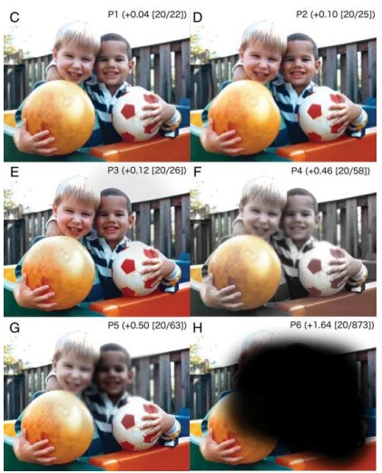
So, I propose a little experiment: let’s take a poll and see which one of those photos is closest to what each of us sees. I am closest to G.
If we get more than six people I will write it up. I suspect the Macular Degeneration Partnership would publish for us. We might be able to be published researchers!
And in other news, when I was reading that article on using a membrane to support stem cell-derived RPEs, I came upon this line: The ELM is a structure present in the normal retina and absent in the area of disease in patients with geographic atrophy.
Oh, hell! Something I am supposed to have, have apparently lost and I don’t even know what it is! ELM?
The External Limiting Membrane is also called the Outer Limiting Membrane (OLM). But whatever it is called, it appears those of us with GA “ain’t got it” any more. According to an article on the histology of the eye published by Researchgate, the retinal is divided into ten layers. With my limited knowledge – and remember we are not experts, not doctors. We are just curious people with access to WiFi – it would appear to me the first seven layer starting from the inside are nearly all some sort of neuron or piece of a neuron. The eighth layer is the ELM/OLM and creates a “junction between Muller cells and photoreceptors.” What else does it do? No clue. I looked at a dozen articles and not one said what the primary purpose is. What they all said was degradation of the ELM/OLM layer is predictive of vision loss.
So where we are knowledge-wise appears to be we with GA have lost our ELM/OLM layer which is somehow crucial for good vision. The article about using the polymer membrane to support the transplanted cells suggested their procedure promoted growth of something that resembled the external limiting membrane and that their procedure might “promote restoration of retinal architecture”. So maybe someday we could get it back???? Who knows.
Anybody know anything about this? Anybody have more questions? Still searching here.
“Trust those who seek the truth but doubt those who say they have found it.” – Andre Gide
