What is Myopic Macular Degeneration?

Question: What is Myopic Macular Degeneration which is also called MMD?

Top of diagram shows normal eye & vision. Below, myopic eye and vision.

Myopic Macular Degeneration (MMD) occurs in some people who are severely shortsighted/nearsighted/myopic. In early myopia, which is very common, the shape of the eye changes from that of a baseball to that of a football. When that happens, the distance from the front of the eye to the retina in the back is longer which can cause problems focusing. At that stage, eyeglasses can help.

When it is severe, the elongation of the eye thins all areas of the retina including the macula. This thinning can cause retinal cells to atrophy which leads to loss of central vision. Severe myopia is called high myopia which can become pathological myopia. If it includes the macula, it’s called Myopic Macular Degeneration, MMD, Myopic MD, Myopic Maculopathy, or degenerative myopia. Many of these terms are used interchangeably.

Potential Problems from Pathological Myopia

The thinning affects the entire retina and can cause retinal tears, retinal detachment, cataracts, and glaucoma.

Myopic Macular Degeneration – CNV

Sometimes the thinning of the retina causes new, unwanted blood vessels to grow from the choroid (normal blood supply to the retina) to the area of the photoreceptors which give us sight. This is called CNV or Myopic CNV or Myopic Choroidal Neovascularization (that’s what CNV means). These new blood vessels are fragile and can leak blood or fluid.  It’s what happens in wet AMD and is sometimes called wet MMD.

You can find out more in the articles Myopic Macular Degeneration and What is Myopic Macular Degeneration?

MMD and AMD

Similarities
Where they are different is the cause:
    • MMD is caused by the action of the eyeball changing from an oval shape to a football shape. That ‘mechanical’ action causes the retina to thin including the area of the macula.
    • AMD is caused by some things we know (genes, age, lifestyle) but some things we don’t know yet.

Treatment

In MMD, AMD, diabetic retinopathy, other types of macular degeneration, when there is a build up of a protein called VEGF, the tiny blood vessels below the macula start to grow toward the RPEs & photorecepters which are both necessary for vision. VEGF is actually a good thing elsewhere in the body because it promotes healing. Not in the retina. This process is called CNV for Choroidal NeoVascularization
and can occur in any form of MD and other retinal diseases. CNV is treated with anti-VEGF injections. 
The word ‘wet’ – some people call CNV in MMD ‘wet’ MMD. In medical language, dry AMD is atrophic and wet is neovascular or exudative. Atrophic means ‘atrophied’ or ’tissue that has atrophied.’ Exudative refers to ‘exudate’ which is the fluid or blood that comes out of blood vessels. It’s because of the exudate/fluid/blood that the term wet AMD or wet MMD is used.

Supplements

The AREDS2-based supplements have not been tested in those with MMD because of the differences in causes. AMD is a disease process, whereas MMD is from a mechanical change in the shape of the eyeball.
Some people with MMD take lutein & zeaxanthin supplements and eat foods high in them because they build up the pigment in the macula which is thinned in MMD. I cannot, however, find any research to prove that this is of benefit.

Living and Thriving with MMD

Here is an interview with Dr. Bonnielin Swenor, from Wilmer Eye Institute at Johns Hopkins whose diagnosis of and journey with Myopic Macular Degeneration led her to her current position at Wilmer as Director of the Johns Hopkins University Disability Health Research Center and Associate Professor of Ophthalmology.

Research

The area of research of the most interest to those with vision loss from any ocular disease is that of stem cells being used to replace failing or dead retina cells. Most of the current research is focused on those with AMD because there are more people with AMD than MMD.

The best way to keep updated on all MMD research is to join the Facebook group ‘Myopic Macular Degeneration : Patients For a Cure’ (link is below). They are actively monitoring all research that is about MMD or could be applied to MMD especially that using stem cells. They were able to set up an online discussion with Professor Pete Coffey of the London Project to Cure Blindness. This is the video recording of that discussion: ‘Myopic macular degeneration and how to find a cure.’

Facebook Groups

If you have MMD, I welcome you to join our Facebook group where you can learn about the disease and how to manage your life with a visual impairment (if you have one). Most of our members do have AMD, but we have members with MMD as well. There are other groups specific to MMD (I don’t belong to the first one list, but I do belong to the second one):


go back to frequently asked questions


Highlight: What is Myopic Macular Degeneration?

There are quite a few eye diseases that cause degeneration of the macula. The most common forms of macular degeneration are defined by age, at least in part:  1) Age-Related MD (AMD or ARMD) and 2) Juvenile MD (JMD) – several inherited and rare diseases that affect children and young adults including Stargardt’s Disease, Best Disease, and juvenile retinoschisis.

There is another form of macular degeneration called Myopic Macular Degeneration or Myopic Maculopathy.

What is myopia?

If you have trouble seeing things far away like road signs, your eye doctor may have said that means you are nearsighted because you can see things near better than far.   Some people call it being shortsighted.  The medical/optical term is myopia.
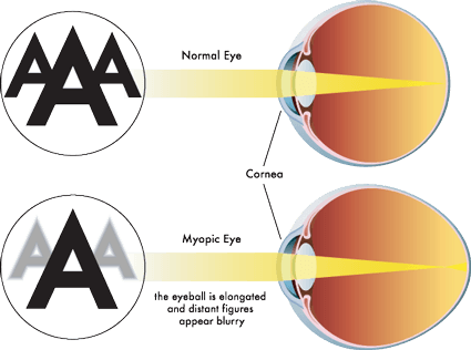
Top of diagram shows normal eye & vision. Below, myopic eye and vision.

Myopia occurs when the eyeball is too long relative to the focusing power of the cornea and lens of the eye (see diagram above). This causes light rays to focus at a point in front of the retina, rather than directly on its surface.   It can also be caused by the cornea and/or lens being too curved for the length of the eyeball. In some cases, myopia is due to a combination of these factors. Click here for more information.

There are different degrees of myopia: mild, moderate and high (also called pathological myopia).   The higher the degree, the higher chance of the degeneration of the macula.  Click here for some other risks for high myopia including retinal detachment, macular detachment, glaucoma and macular holes.

Myopic Macular Degeneration

People with high myopia/pathological myopia are at risk for damage to the macula from the stretching of it as the eyeball grows throughout life.  Here’s a good description of what happens: “As the eyeball grows and stretches, it may also cause an area of atrophy and/or cracks in the layers under the retina. These cracks can serve as conduits for abnormal blood vessels to grow under the retina. These vessels can hemorrhage and scar which is similar to what happens in wet macular degeneration. The onset of the nets of abnormal blood vessels, called Fuch’s spots, often occur in the 4th to 6th decades of life. Approximately 5% of pathological myopia patients develop Fuch’s spots, which lead to damage in the macular region of the eye and a subsequent loss of central vision.” From http://www.lowvision.org/Pathological%20Myopia.htm

Not all people who are myopic get Myopic Macular Degeneration.

Click here for an explanation of how the atrophy can occur as well as how the blood vessels grow into the macula.

The symptoms are similar to AMD & JMD as well.  Click here to learn more about them.  This article will take you to stories of people who have Myopic MD.

Click here for a first-hand account of the disease from a person with it.

Other links:

Myopic Macular Degeneration

American Academy of Ophthalmology Diagnosis and Treatment of CNV in Myopic Macular Degeneration

Degenerative Myopia

Treatment Patterns for Myopic Choroidal Neovascularization in the United States, article published July 2017 edition of AAO Journal (AAO is American Academy of Ophthalmologists).