Highlight: Here’s a GREAT website especially for those with wet AMD

Lin/Linda here: Every once in a while I find a website and/or Facebook page that stands out.  Here’s one of those.

The website and Facebook page are called The Science of AMD: Our vision is to save your vision.  It is presented by the Amgiogenesis Foundation. Their headquarters are in Cambridge, Massachusetts.


Click here to go to the website. From there, you can connect to Facebook, Twitter or YouTube using icons in the upper right corner.


What is angiogenesis? From the website: “Angiogenesis is the process used by the body to grow blood vessels. In healthy adults, normal angiogenesis occurs in healing wounds and reproduction, but in all other situations, it is abnormal.”

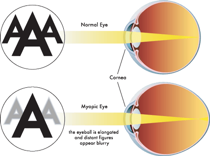
It’s what causes wet AMD: “Wet AMD is caused by abnormal angiogenesis, when new vessels grow under the macula, disrupting the central region of the retina. These new blood vessels bleed and leak fluid, causing the macula to bulge or lift up from its normally flat position, impairing central vision. If left untreated, scar tissue can form, and central vision is irreversibly lost.” 

What’s so special about the website?
  • From a design standpoint, you can change the size of the font and the color of the font & background, you can choose a version of the site in any of 7 languages as depicted by flags, it’s easy to navigate.
  • Format of content includes printed text, videos, audio, graphics, PDF files and more.
  • This is not just for the US, there are resources available for other countries as well.
What information can I find there?

There’s a menu with Learn, Treat, Resources, Connect, About, Donate.  I suggest you start at Learn!   The emphasis is on how angiogenesis causes wet AMD and what can be done to treat it.

OK, now go and explore! Let me know what you think!

Highlight: What is Myopic Macular Degeneration?

There are quite a few eye diseases that cause degeneration of the macula. The most common forms of macular degeneration are defined by age, at least in part:  1) Age-Related MD (AMD or ARMD) and 2) Juvenile MD (JMD) – several inherited and rare diseases that affect children and young adults including Stargardt’s Disease, Best Disease, and juvenile retinoschisis.

There is another form of macular degeneration called Myopic Macular Degeneration or Myopic Maculopathy.

What is myopia?

If you have trouble seeing things far away like road signs, your eye doctor may have said that means you are nearsighted because you can see things near better than far.   Some people call it being shortsighted.  The medical/optical term is myopia.

Top of diagram shows normal eye & vision. Below, myopic eye and vision.

Myopia occurs when the eyeball is too long relative to the focusing power of the cornea and lens of the eye (see diagram above). This causes light rays to focus at a point in front of the retina, rather than directly on its surface.   It can also be caused by the cornea and/or lens being too curved for the length of the eyeball. In some cases, myopia is due to a combination of these factors. Click here for more information.

There are different degrees of myopia: mild, moderate and high (also called pathological myopia).   The higher the degree, the higher chance of the degeneration of the macula.  Click here for some other risks for high myopia including retinal detachment, macular detachment, glaucoma and macular holes.

Myopic Macular Degeneration

People with high myopia/pathological myopia are at risk for damage to the macula from the stretching of it as the eyeball grows throughout life.  Here’s a good description of what happens: “As the eyeball grows and stretches, it may also cause an area of atrophy and/or cracks in the layers under the retina. These cracks can serve as conduits for abnormal blood vessels to grow under the retina. These vessels can hemorrhage and scar which is similar to what happens in wet macular degeneration. The onset of the nets of abnormal blood vessels, called Fuch’s spots, often occur in the 4th to 6th decades of life. Approximately 5% of pathological myopia patients develop Fuch’s spots, which lead to damage in the macular region of the eye and a subsequent loss of central vision.” From http://www.lowvision.org/Pathological%20Myopia.htm

Not all people who are myopic get Myopic Macular Degeneration.

Click here for an explanation of how the atrophy can occur as well as how the blood vessels grow into the macula.

The symptoms are similar to AMD & JMD as well.  Click here to learn more about them.  This article will take you to stories of people who have Myopic MD.

Click here for a first-hand account of the disease from a person with it.

Other links:

Myopic Macular Degeneration

American Academy of Ophthalmology Diagnosis and Treatment of CNV in Myopic Macular Degeneration

Degenerative Myopia

Treatment Patterns for Myopic Choroidal Neovascularization in the United States, article published July 2017 edition of AAO Journal (AAO is American Academy of Ophthalmologists).


News: Summary of AMD Research & Developments for past 12 months – June 2016

Here’s an excellent summary of the research and developments in the field of AMD.  It’s done by Dan Roberts of mdsupport.org which is a site I highly recommend for both information and support.

Click here for the audio with slides presentation. You don’t have to be able to see the slides, the audio is very clear.

Click here for a transcript of the presentation.


Highlight: What is Advanced Macular Degeneration?

About 10 years ago, my elderly father was diagnosed with dry AMD and then later we were told it was ‘advanced’ and that he had ‘geographic atrophy’.  At the time, I didn’t understand what that was.

Here’s an excellent article about what those terms really mean.  It’s on the great website WebRN: Macular Degeneration.  If you haven’t looked at it, I recommend that you do after you read this article.

Advanced AMD & Geographic Atrophy