Highlight: Can wet AMD turn to dry AMD? 10/2/2018

We have a GREAT Facebook group (as I’m typing, there are 1,898 members, but that will change shortly since I add at least a few new members every day).  They share information and ask me questions and also ask questions of other members.  That’s one of the reasons that I started the group: to encourage discussion.

A recent discussion of what happens after anti-VEGF injections brought up the confusing use of the word “dry” when talking about wet AMD.  Doctors will often tell patients, “Your eye is dry,” or, “Your AMD has dried up.”  One of the members said that’s what she was told. Another member asked, “Can wet AMD turn to dry AMD?” Because of the confusing use of the word “dry”, other members said, “Yes!” It turns out that they were mostly referring to the correct use of the word, but I thought it a good time to clarify this.

When the doctor tells you that your wet AMD is now “dry”
Click on image to see a larger version of this illustration.

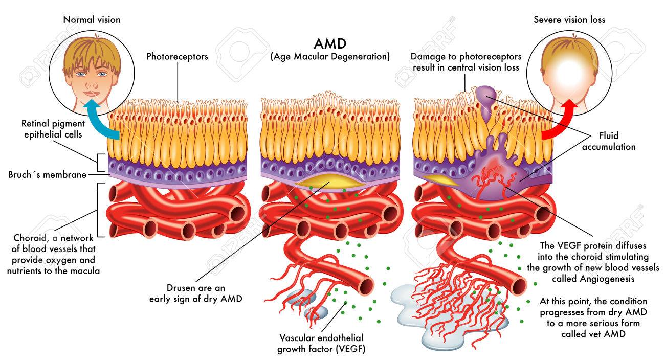
In the diagram, going from left to right is the disease PROCESS from no AMD to dry then to wet AMD. Not everyone gets to the last & wet stage (10-15% of those who have AMD), but if they do, the disease process continues. If the injection was successful, the fluid that was leaking has “dried up.” Do NOT let down your guard thinking you have dry AMD and stop checking your vision or going to the eye specialist. Unfortunately, there is no way to reverse the disease process yet.

Another way to view the process
Click on the image above to for a larger version of the diagram.

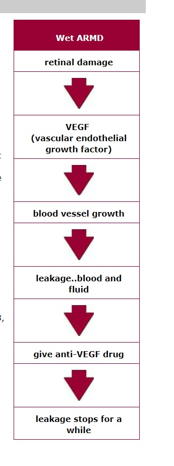
This diagram is from the most comprehensive page on wet AMD I have found. Some of it is highly technical, but it should give you a sense of what happens when the disease process of AMD progresses to wet.  There are no arrows pointing up at the bottom of the diagram. The fluid from wet AMD can ‘dry up’ – that’s the objective – but the disease process does not go back to an earlier stage. Some people will say that if the macula is “dry”/”dried up”, the disease goes “dormant.” Regardless of the terminology, the disease process cannot be reversed yet. The injections treat the symptoms that affect visual acuity.

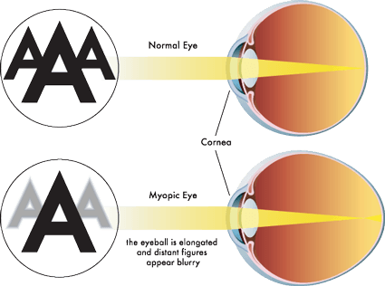
Another name for wet AMD is CNV which is Choroidal NeoVascularization (CNV is also used in other forms of macular degeneration).  The choroid is where the blood vessels are below Bruch’s Membrane and the RPEs and photoreceptors (see the illustration at the top of the page).  Neo means “new” and vascularization is abnormal or excessive formation of blood vessels.

Do you have questions about wet AMD?

Of course, we’d love if you’d join the Facebook group, but we know that some of you have chosen not to for various reasons – and that’s okay.  If you have questions, please put them in the ‘leave a reply’ section, and I’ll answer them as soon as I can.


UPDATED: Are there eye drops instead of injections to treat wet AMD?

The short answer to that question: not yet.  There is a major problem in developing eye drops for AMD.  One article states it as “Ophthalmic drugs have traditionally been administered topically, which in general provides therapeutic levels to the anterior (front) chamber of the eye but not to the posterior (back) segment. Therefore, topical administration of drugs has been largely infeasible for posterior segment diseases such as AMD and diabetic macular oedema.  In contrast, intravitreous injection [for wet AMD] provides direct delivery to the posterior segment and allows therapeutic levels to be attained.”

Here are the studies so far (there is other research that I’m not including that is currently only being done with animals).  Keep in mind that there are four phases to clinical trials.  Click here for more information about clinical trials.

  • PAN-90806/Panoptica: topical Lucentis
    • clinicaltrials.gov Phase I completed
    • “A phase I/II trial of the next generation of the formulation is set to begin in the first and second quarters of 2017.” Click here for an article.
  • OHR-102 (originally “Squalamine”): squalamine has been shown to interrupt and reverse the process of angiogenesis; used in conjunction with Lucentis injections, goal is to reduce number of injections or eliminate them.
    • clinical trials phase III not recruiting subjects,  completed in mid-2019.
    • click here for an article.
    • Temporary suspension of clinical trial phase 2, not due to safety issues.  Going forward with phase III.  “Effectiveness: Unfortunately, Squalamine Eye Drops failed to decrease the average number of Lucentis injections required by the study participants. This was the primary goal of the clinical trial and the result was disappointing, both to researchers and to people with wet AMD, who were hoping that Squalamine Eye Drops could possibly reduce, or even eliminate, the need for eye injections.”
  • CPP: cell-penetrating peptide (CPP) can be used to deliver therapeutically relevant doses of ranibizumab (Lucentis) and bevacizumab (Avastin)
    • University of Birmingham, England
    • click here for an article.
    • “This is exciting for both patients and practitioners,” said Dr. Jayanth Sridhar, assistant professor of clinical ophthalmology at the Bascom Palmer Eye Institute. “But this was a preliminary study in animal eyes only. Further research must be undertaken in human subjects first to establish safety, and then to establish effectiveness. Still, this study offers at least a glimmer of hope that in the coming years we may see a topical drop option emerge to supplement or replace injections.”
  • OC-118: “solubilizing nanoparticle” technology
    • Article Jan. 6. 2018 says “This technology, the company hopes, can boost the drug’s ability to reach the front and the back of the eye — something that previous drugs haven’t achieved unless delivered via needle to the eye.” It is currently being looked at to treat Diabetic Macular Edema (DME). I couldn’t find anything to confirm or deny that it could be used to treat AMD.

Updated & verified 1/8/2018

Highlight: Here’s a GREAT website especially for those with wet AMD

Lin/Linda here: Every once in a while I find a website and/or Facebook page that stands out.  Here’s one of those.

The website and Facebook page are called The Science of AMD: Our vision is to save your vision.  It is presented by the Amgiogenesis Foundation. Their headquarters are in Cambridge, Massachusetts.


Click here to go to the website. From there, you can connect to Facebook, Twitter or YouTube using icons in the upper right corner.


What is angiogenesis? From the website: “Angiogenesis is the process used by the body to grow blood vessels. In healthy adults, normal angiogenesis occurs in healing wounds and reproduction, but in all other situations, it is abnormal.”

It’s what causes wet AMD: “Wet AMD is caused by abnormal angiogenesis, when new vessels grow under the macula, disrupting the central region of the retina. These new blood vessels bleed and leak fluid, causing the macula to bulge or lift up from its normally flat position, impairing central vision. If left untreated, scar tissue can form, and central vision is irreversibly lost.” 

What’s so special about the website?
  • From a design standpoint, you can change the size of the font and the color of the font & background, you can choose a version of the site in any of 7 languages as depicted by flags, it’s easy to navigate.
  • Format of content includes printed text, videos, audio, graphics, PDF files and more.
  • This is not just for the US, there are resources available for other countries as well.
What information can I find there?

There’s a menu with Learn, Treat, Resources, Connect, About, Donate.  I suggest you start at Learn!   The emphasis is on how angiogenesis causes wet AMD and what can be done to treat it.

OK, now go and explore! Let me know what you think!

UPDATED: Are there eye drops instead of injections to treat wet AMD?

The short answer to that question: not yet.  There is a major problem in developing eye drops for AMD.  One article states it as “Ophthalmic drugs have traditionally been administered topically, which in general provides therapeutic levels to the anterior (front) chamber of the eye but not to the Continue reading “UPDATED: Are there eye drops instead of injections to treat wet AMD?”

Highlight: What is Myopic Macular Degeneration?

There are quite a few eye diseases that cause degeneration of the macula. The most common forms of macular degeneration are defined by age, at least in part:  1) Age-Related MD (AMD or ARMD) and 2) Juvenile MD (JMD) – several inherited and rare diseases that affect children and young adults including Stargardt’s Disease, Best Disease, and juvenile retinoschisis.

There is another form of macular degeneration called Myopic Macular Degeneration or Myopic Maculopathy.

What is myopia?

If you have trouble seeing things far away like road signs, your eye doctor may have said that means you are nearsighted because you can see things near better than far.   Some people call it being shortsighted.  The medical/optical term is myopia.

Top of diagram shows normal eye & vision. Below, myopic eye and vision.

Myopia occurs when the eyeball is too long relative to the focusing power of the cornea and lens of the eye (see diagram above). This causes light rays to focus at a point in front of the retina, rather than directly on its surface.   It can also be caused by the cornea and/or lens being too curved for the length of the eyeball. In some cases, myopia is due to a combination of these factors. Click here for more information.

There are different degrees of myopia: mild, moderate and high (also called pathological myopia).   The higher the degree, the higher chance of the degeneration of the macula.  Click here for some other risks for high myopia including retinal detachment, macular detachment, glaucoma and macular holes.

Myopic Macular Degeneration

People with high myopia/pathological myopia are at risk for damage to the macula from the stretching of it as the eyeball grows throughout life.  Here’s a good description of what happens: “As the eyeball grows and stretches, it may also cause an area of atrophy and/or cracks in the layers under the retina. These cracks can serve as conduits for abnormal blood vessels to grow under the retina. These vessels can hemorrhage and scar which is similar to what happens in wet macular degeneration. The onset of the nets of abnormal blood vessels, called Fuch’s spots, often occur in the 4th to 6th decades of life. Approximately 5% of pathological myopia patients develop Fuch’s spots, which lead to damage in the macular region of the eye and a subsequent loss of central vision.” From http://www.lowvision.org/Pathological%20Myopia.htm

Not all people who are myopic get Myopic Macular Degeneration.

Click here for an explanation of how the atrophy can occur as well as how the blood vessels grow into the macula.

The symptoms are similar to AMD & JMD as well.  Click here to learn more about them.  This article will take you to stories of people who have Myopic MD.

Click here for a first-hand account of the disease from a person with it.

Other links:

Myopic Macular Degeneration

American Academy of Ophthalmology Diagnosis and Treatment of CNV in Myopic Macular Degeneration

Degenerative Myopia

Treatment Patterns for Myopic Choroidal Neovascularization in the United States, article published July 2017 edition of AAO Journal (AAO is American Academy of Ophthalmologists).


Highlight: Does wet AMD start as dry?

There are differing opinions and interpretations of what those in the field of macular degeneration have learned.  The links below certainly don’t represent all views, of course, but are an example of some of what you will find.

The important point is that it is CRITICAL to get your eyes examined as recommended by your doctor and to monitor your vision between exams. Click here to find out how to do that.

From: http://www.webrn-maculardegeneration.com/macular-degeneration-stages.html

“Advanced AMD is either a break-down of light sensitive cells and supporting tissue in the central retinal area (advanced dry form), or the development of abnormal and fragile blood vessels under the retina (wet form).”

From: https://www.macular.org/dry-vs-wet-macular-degeneration

“About ten percent of all cases of Age-related Macular Degeneration become “Wet” AMD (typically a person has dry AMD first and progresses toward wet).”

From: https://nei.nih.gov/health/maculardegen/armd_facts

“There are three stages of AMD defined in part by the size and number of drusen under the retina. It is possible to have AMD in one eye only, or to have one eye with a later stage of AMD than the other.

Early AMD. Early AMD is diagnosed by the presence of medium-sized drusen, which are about the width of an average human hair. People with early AMD typically do not have vision loss.

Intermediate AMD. People with intermediate AMD typically have large drusen, pigment changes in the retina, or both. Again, these changes can only be detected during an eye exam. Intermediate AMD may cause some vision loss, but most people will not experience any symptoms.

Late AMD. In addition to drusen, people with late AMD have vision loss from damage to the macula. There are two types of late AMD:

In geographic atrophy (also called dry AMD), there is a gradual breakdown of the light-sensitive cells in the macula that convey visual information to the brain, and of the supporting tissue beneath the macula. These changes cause vision loss.

In neovascular AMD (also called wet AMD), abnormal blood vessels grow underneath the retina. (“Neovascular” literally means “new vessels.”) These vessels can leak fluid and blood, which may lead to swelling and damage of the macula. The damage may be rapid and severe, unlike the more gradual course of geographic atrophy. It is possible to have both geographic atrophy and neovascular AMD in the same eye, and either condition can appear first.”

From: http://www.avruc.com/macular-degeneration.php

“The macula is the central part of the retina and allows us to read fine print clearly and see colors vividly. It is this area of the retina that deteriorates in ARMD. There are 2 forms of ARMD. Everyone who has macular degeneration starts out with the dry type and 20% progress to the wet type over the course of a lifetime. “

From: http://www.youreyes.org/eyehealth/macular-degeneration

“Can the dry form turn into the wet form?
Yes. All people who had wet form AMD had the dry form first. 

The dry form can advance and cause vision loss without turning into the wet form. The dry form also can suddenly turn into the wet form, even during early stage AMD. There is no way to tell if or when the dry form will turn into the wet form.

Can advanced AMD be either the dry form or the wet form?
Yes. Both the wet form and the advanced dry form are considered advanced AMD. Vision loss occurs with either form. In most cases, only advanced AMD can cause vision loss. 

People who have advanced AMD in one eye are at especially high risk of developing advanced AMD in the other eye.”

Please see your doctor for regular exams and between exams, check your vision at home.